
Chest CT Can Measure Bone Density and Warn of Brain Aging Risk: Latest Research Reveals Link Between Skeletal and Cognitive Decline
 Scan to share
Chest computed tomography (CT), routinely used to screen for lung lesions, may hold important clues for assessing brain health. A research team from Johns Hopkins University in the United States explored approaches to using existing chest imaging to assess bone health and link it to brain aging, with the related findings published in September 2026 in the authoritative journal Radiology. The study showed that subjects with lower thoracic vertebral bone density experienced faster cognitive decline during subsequent follow-up, and specific white matter microstructural changes also showed associated degeneration.
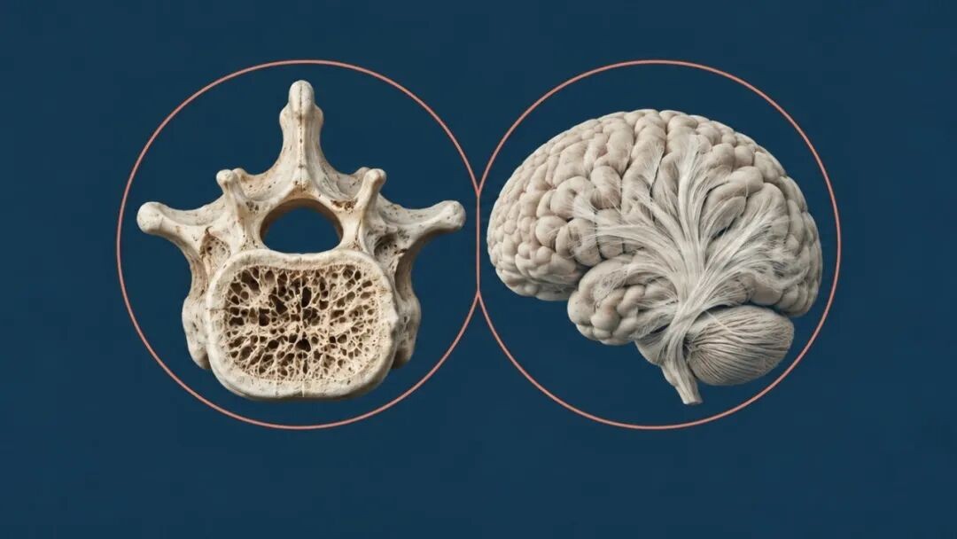
Chest CT is widely used in lung cancer screening and pulmonary nodule follow-up, and the images simultaneously contain clear thoracic vertebral structures. Dr. Sara Momtazmanesh, the study's first author, pointed out that such images contain a large amount of additional clinical value that has not yet been fully utilized. Using the deep learning anatomical segmentation tool TotalSegmentator, researchers were able to automatically locate the first through tenth thoracic vertebrae, algorithmically remove the dense outer cortical bone, and precisely quantify three-dimensional volumetric bone density of the inner spongy trabecular bone, eliminating the burden of additional dedicated bone density examinations for patients.
To investigate the link between bone density and brain degeneration, the research team relied on the Multi-Ethnic Study of Atherosclerosis (MESA) cohort in the United States, enrolling 715 subjects with a median age of 69 and conducting approximately 5 years of follow-up. Researchers quantified the subjects' baseline thoracic vertebral bone density and compared it with subsequent neurocognitive assessment scores and brain magnetic resonance imaging (MRI) data, with more than 400 subjects having complete white matter tract imaging.
Among neural structures, gray matter dominates information processing, while white matter serves as the information pathways for efficient transmission of neural signals between brain regions. Researchers assessed the degree of microstructural degeneration of nerve fibers by detecting white matter hyperintensities and water molecule diffusion metrics on MRI. The results showed that individuals with lower baseline thoracic vertebral bone density had a more significant decline in overall cognitive scores, with white matter structural alterations particularly prominent in the corpus callosum and the anterior limb of the internal capsule. These two brain regions serve as key hubs for communication between the left and right cerebral hemispheres and neural network transmission, directly participating in the regulation of attention and executive function.
The research team emphasized that the current findings reveal a statistical correlation between bone density and brain health decline, rather than a direct causal relationship. Senior author Professor Shadpour Demehri analyzed that shared systemic physiological mechanisms such as insulin resistance, lipid metabolism disorders, and menopause-related hormonal changes may simultaneously accelerate bone loss and the aging process of cerebrovascular and neural tissues.
The core value of this achievement lies in "one scan, multiple uses," namely deeply mining potential health risk signals from existing routine examination data, rather than recommending that the public simply undergo additional radiological examinations for brain health screening. Currently, the algorithm and prediction model remain at the research stage, and future clinical validation is still needed regarding cross-device compatibility, clinical generalizability under different scanning parameters, and the independent predictive weight it can provide beyond traditional aging indicators.
Disclaimer: Information republished from partner media, institutions or other websites is provided for reference and communication purposes only. It does not imply endorsement of its views or verification of its accuracy. Please contact us if any content infringes rights or requires correction.