
PETRA III Synchrotron Radiation Study Reveals New Clues to Aging Mechanisms of the Uterine Broad Ligament
 Scan to share
Austrian and German research teams recently used the PETRA III facility at the German Electron Synchrotron (DESY) to conduct X-ray analysis of the broad ligament, an important ligament supporting the uterus, providing new biomechanical clues for understanding the mechanisms of pelvic organ prolapse. The findings were published in *Acta Biomaterialia*.
Pelvic organ prolapse is a common but poorly understood issue in women's health, affecting approximately 40% of women, and predominantly occurring in the latter half of life. Medical researchers have previously speculated that age-related loosening of the ligaments surrounding the uterus may be a significant contributing factor to prolapse. The broad ligament connects the uterus to the pelvic wall, and its strength and elasticity primarily derive from collagen, making the question of whether collagen degrades with age a central focus for the research team.
A team led by researchers from the Medical University of Graz and the University Medical Center Hamburg-Eppendorf selected uterine broad ligament samples from women aged 18 to 89 years and examined them using small-angle X-ray scattering at the P03 beamline of PETRA III. The researchers used sub-millimeter ligament tissue sections and applied biaxial tensile stress to the samples during the experiments to observe changes in collagen structure under load.
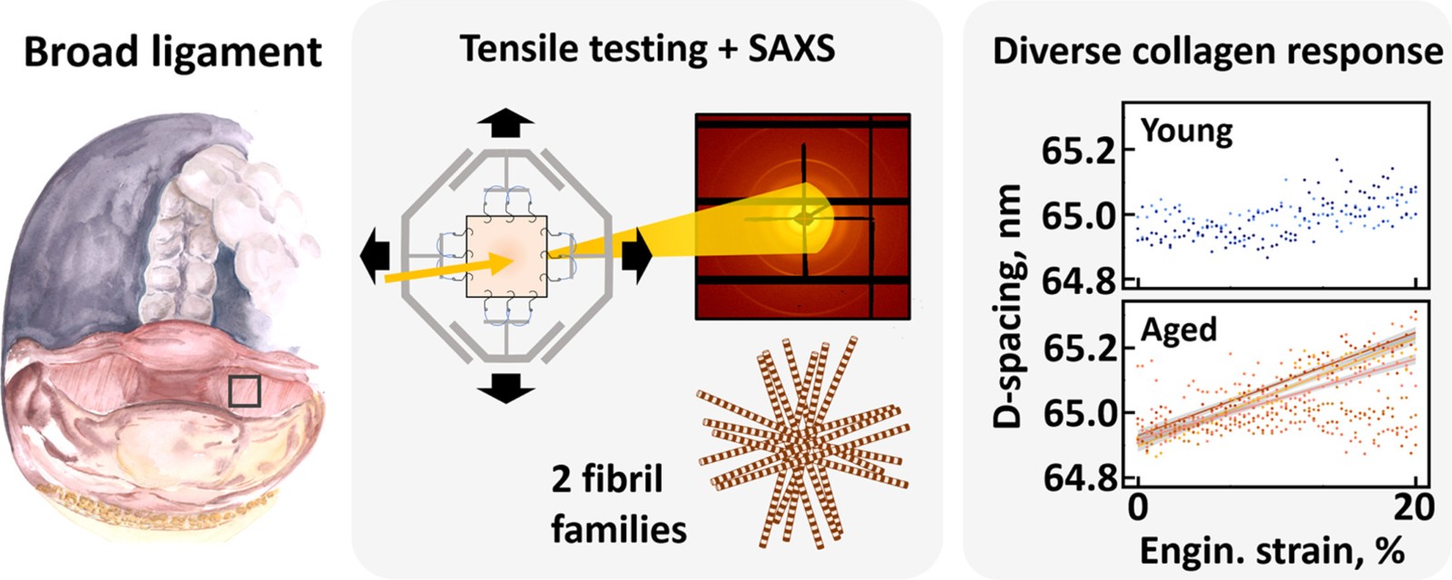
Experimental overview and results: A: Adjacent uterine mesometrium samples were used for small-angle X-ray scattering (B) histological analysis under different tensile testing protocols (C) behavior during continuous tensile testing up to 20%: young tissue samples showed no significant increase, while 3 of 7 aged tissue samples showed a marked and significant increase in collagen spacing ("d-spacing") (Graphical abstract from the original publication under CC BY 4.0 license).
The experimental results showed no significant overall differences in collagen between broad ligaments from young and older women. The research team concluded that aging alone may not directly lead to structural degradation of broad ligament collagen. However, in some samples from older women, subtle nanoscale changes in collagen were observed; during continuous tensile testing up to 20%, 3 of 7 aged tissue samples showed a significant increase in collagen spacing.
The researchers stated that these findings indicate that the behavior of broad ligament collagen does not exhibit a clear age-related pattern, but subtle structural changes in some samples may be associated with disease development and progression. The results contribute to a better understanding of the mechanical behavior of uterine supporting tissues and may also provide a reference for future improvements in pelvic floor disease treatment strategies and for evaluating the quality of surgical tissues.
Disclaimer: Information republished from partner media, institutions or other websites is provided for reference and communication purposes only. It does not imply endorsement of its views or verification of its accuracy. Please contact us if any content infringes rights or requires correction.