
University of Tokyo and University of York Successfully Validate Positronium Ratio Imaging Principle for the First Time
 Scan to share
A research team from the Graduate School of Engineering at the University of Tokyo and the University of York in the UK established a novel geometric imaging principle utilizing positron three-photon decay. Using a gamma-ray measurement system comprising scintillators and silicon photomultipliers (SiPMs), along with the existing PET radiopharmaceutical ^18F-FDG, they successfully visualized the positronium three-photon decay ratio for the first time, completing the proof-of-principle for Positronium Ratio Imaging (PRI). The findings were published in *Communications Physics*.
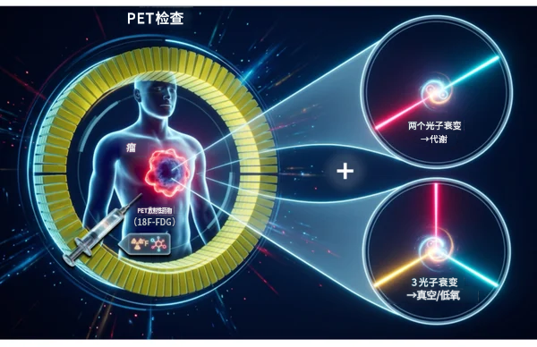
Current clinical nuclear medicine PET examinations primarily utilize gamma rays generated from two-photon annihilation between positrons and electrons to visualize drug accumulation in the body. While this method provides quantitative information on metabolism or drug uptake, it offers limited insight into microenvironmental features such as tissue hypoxia, which are closely related to disease malignancy. In recent years, researchers have attempted to obtain additional tissue information through the average lifetime of positronium formed during positron annihilation, but this approach typically requires the introduction of special nuclides or agents, limiting its practical application.
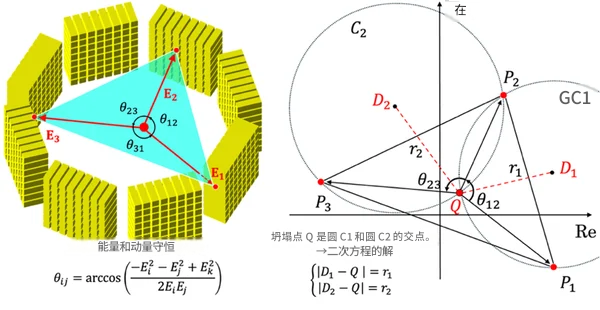
Schematic diagram of three-photon imaging principle
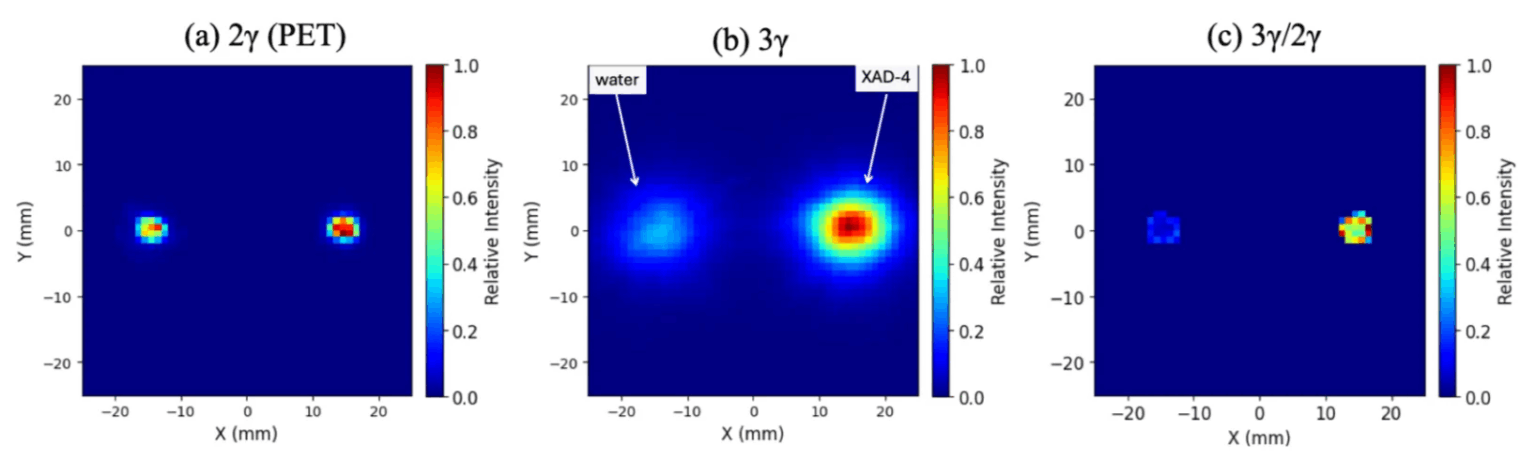
Imaging results when water and porous material (XAD-4) are mixed with 18F-FDG
This study focused on the ratio between positronium three-photon decay and two-photon decay. The research team leveraged the geometric symmetry in the energy and momentum conservation relationships satisfied by three-photon decay, simplifying the originally complex image reconstruction problem into an analytically solvable quadratic equation. Concurrently, the team developed a detection system compatible with clinical PET equipment, employing high-energy-resolution HR-GAGG scintillator arrays and SiPM photodetector arrays to meet the detection performance requirements of three-photon imaging.
In the experiments, the researchers mixed water and the porous material XAD-4 with ^18F-FDG respectively and performed imaging. The results showed a significant increase in the positronium ratio in the porous material, validating the capability of PRI technology to distinguish different microenvironments. The research team stated that this achievement demonstrates the possibility of obtaining higher-level tissue microenvironment information using existing clinical PET agents without developing new PET radiopharmaceuticals.
Potential applications of PRI technology include non-invasive observation of microenvironmental features such as void size and hypoxic status within biological organisms or materials. Particularly in tumor diagnosis and treatment, tissue hypoxia is closely associated with cancer malignancy and treatment resistance; if quantitative assessment can be achieved, it is expected to facilitate earlier formulation of appropriate treatment strategies. The technology is also considered potentially useful for early research and diagnostic exploration of diseases such as Alzheimer's disease.
Furthermore, the research team believes that PRI is highly compatible with the theranostics technology currently developing in the nuclear medicine field. For example, combining nuclides with both diagnostic and therapeutic properties, such as copper-64 (^64Cu), could enable quantification of tumor metabolism and hypoxic environment while conducting PET imaging and PRI assessment, providing additional evidence for personalized treatment.
Disclaimer: Information republished from partner media, institutions or other websites is provided for reference and communication purposes only. It does not imply endorsement of its views or verification of its accuracy. Please contact us if any content infringes rights or requires correction.