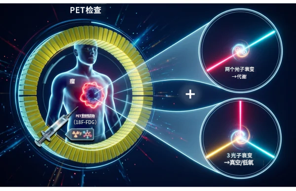
University of Tokyo and University of York Successfully Validate Positronium Ratio Imaging Principle for the First Time
A research team from the Graduate School of Engineering at the University of Tokyo and the University of York in the UK established a novel geometric imaging principle utilizing positron three-photon decay. Using a gamma-ray measurement system comprising scintillators and silicon photomultipliers (SiPMs), along with the existing PET radiopharmaceutical ^18F-FDG, they successfully visualized the positronium three-photon decay ratio for the first time, completing the proof-of-principle for Positronium Ratio Imaging (PRI). The findings were published in *Communications Physics*. Current clinical nuclear medicine PET examinations primarily utilize gamma rays generated from two-photon annihilation between positrons and electrons to visualize drug accumulation in the body. While this method provides quantitative information on metabolism or drug uptake, it offers limited insight into microenvironmental features such as tissue hypoxia, which are closely related to disease malignancy. In recent years, researchers have attempted to obtain additional tissue information through the average lifetime of positronium formed during positron annihilation, but this approach typically requires the introduction of special nuclides or agents, limiting its practical application.
2026-08-12