
Russian Researchers Propose New Method for Preparing Iron-59 Magnetite Nanoparticles, Potentially Advancing Nuclear Medicine Diagnostics and Therapy
 Scan to share
Russian researchers have developed a new method for preparing iron-59-labeled magnetite nanoparticles, which is expected to provide a new material preparation pathway for medical applications such as magnetic resonance imaging, single-photon emission computed tomography, and radionuclide therapy.
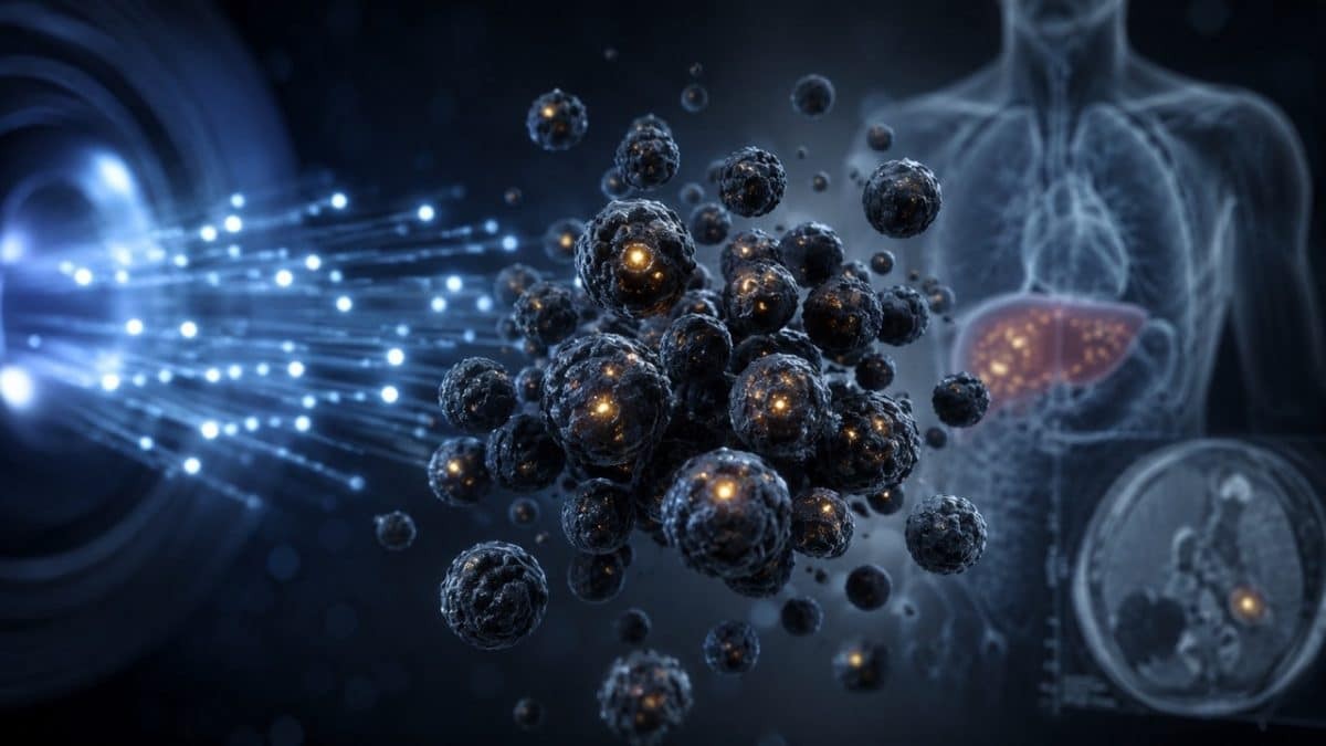
According to reports, researchers from the Vernadsky Institute of Geochemistry and Analytical Chemistry and the Karpov Institute of Physical Chemistry, both under the Russian Academy of Sciences, have proposed synthesizing iron-59 magnetite nanoparticles using the sol-gel method. The relevant findings have been published in the journal Applied Radiation and Isotopes.
The sol-gel method is a chemical approach for preparing nanomaterials, with the core process involving the conversion of a liquid colloidal solution into a gel-like network. In this study, high-purity iron sulfate was used to synthesize magnetite nanoparticles, with a focus on reducing harmful radionuclide impurities that may be generated during thermal neutron activation, particularly avoiding radioactive chlorine isotope byproducts that could arise from traditional raw materials.
Iron-59 is a radioactive isotope with a half-life of 44.5 days. Magnetite nanoparticles containing iron-59 have attracted attention in the nuclear medicine field in recent years due to their relatively low toxicity and the relatively easy availability of raw material sources. Researchers believe that these nanoparticles exhibit superparamagnetic properties and can serve as contrast agents for magnetic resonance imaging. They are easily magnetized in an external magnetic field but do not remain magnetized for extended periods after the field is removed, and they are less prone to aggregation, thereby helping to improve image quality.
In addition to magnetic resonance imaging, the radiation emitted by iron-59 can also be used for single-photon emission computed tomography imaging. According to available data, this imaging modality has application value in cases where metallic implants in the human body may interfere with conventional magnetic resonance imaging. Meanwhile, iron-59 atoms also release β particles during decay, making it a nanoparticle labeling agent considered for use in tumor therapy research.
Previously, the preparation of magnetite nanoparticles faced challenges such as difficulties in manufacturing and storing raw materials, as well as stringent requirements for particle size control. For example, iron chloride, which was previously used to prepare magnetite particles, is prone to undesirable chemical reactions in air; moreover, if neutron activation methods are employed, radioactive chlorine isotopes may be generated, affecting subsequent material processing.
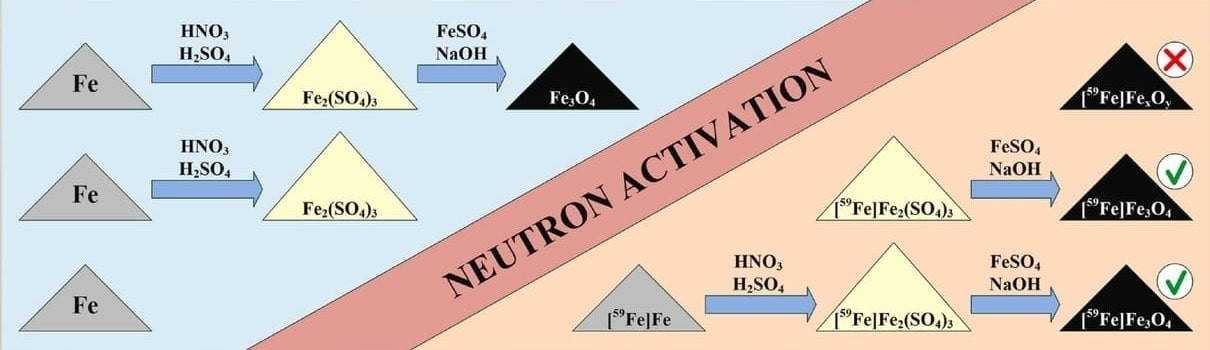
Schematic diagram of the experiment for preparing iron-59-labeled magnetite nanoparticles
In the new method, researchers used high-purity iron sulfate to synthesize nanoparticles via the sol-gel method, yielding particles with an average size of approximately 12 nanometers and the desired superparamagnetic properties. Subsequently, the nanoparticles were placed in the channel of the VVR-ts nuclear reactor in Obninsk, Russia, where iron-59 atoms were generated through thermal neutron activation.
The research results show that synthesizing iron-59-labeled magnetite nanoparticles using activated metallic iron or iron sulfate is highly efficient. The researchers believe that this method nearly eliminates unnecessary byproducts such as radioactive chlorine isotopes, helping to scale up the production of iron-59 nanoparticles without relying on complex and costly processes.
Disclaimer: Information republished from partner media, institutions or other websites is provided for reference and communication purposes only. It does not imply endorsement of its views or verification of its accuracy. Please contact us if any content infringes rights or requires correction.