
South Korean Study Confirms Low-Dose CT Arthrography Reduces Radiation Exposure in Shoulder Examinations
 Scan to share
A research team led by Professor Choi Jeong-ah from the Department of Radiology at Hallym University Dongtan Sacred Heart Hospital in South Korea has recently confirmed that low-dose CT arthrography can reduce patient radiation exposure by approximately 31% while maintaining high diagnostic accuracy, with diagnostic performance comparable to 3T magnetic resonance imaging (3T MRI). The findings have been published in the international musculoskeletal imaging journal *Skeletal Radiology*.
Rotator cuff tears and superior labral anterior-posterior (SLAP) lesions are significant causes of shoulder pain; if not diagnosed and treated in a timely manner, they may lead to arthritis or decreased shoulder joint function. Currently, 3T MRI is commonly used for precise examinations, but it has drawbacks such as high cost, long examination times, and the potential for claustrophobia in some patients; MRI may also be limited for patients with implanted cardiac pacemakers or metal implants. Traditional CT arthrography can serve as an alternative examination method, but radiation exposure has always been a concern.
To evaluate the clinical value of the low-dose protocol, the research team enrolled 60 patients with suspected shoulder diseases, of whom 25 underwent standard-dose CT arthrography and 35 underwent low-dose CT arthrography. The examination results were then compared and analyzed with 3T MRI.
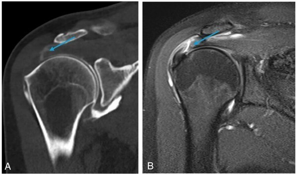
The image shows a comparison of low-dose CT arthrography (left) and standard-dose CT arthrography (right) in a patient with a rotator cuff tear. The low-dose CT arthrography image demonstrates good image quality, with lower noise, higher contrast, and excellent reproducibility in localizing the tear site.
The results showed that the low-dose CT protocol reduced the radiation dose from 4.80 mGy to 3.29 mGy, a decrease of approximately 31%. In terms of image quality, the background noise of the low-dose examination was 11.04 HU, lower than the 16.21 HU of the standard-dose examination, indicating clearer imaging. A comparison of images from a patient with a rotator cuff tear in the study also showed that low-dose CT arthrography performed well in noise control, contrast, and reproducibility of tear site localization.
Diagnostic performance evaluation showed that low-dose CT had a sensitivity of 53.8% and specificity of 100% for diagnosing rotator cuff tears; standard-dose CT had a sensitivity of 40% and specificity of 100%. In diagnosing SLAP lesions, low-dose CT had a sensitivity of 88% and specificity of 80%, compared to standard-dose CT's sensitivity of 93.7% and specificity of 66.7%, indicating comparable overall diagnostic capability.
Choi Jeong-ah stated that the study demonstrates that low-dose CT arthrography can serve as an alternative that balances patient safety and diagnostic accuracy, helping to alleviate concerns about radiation exposure. She noted that the team will continue to promote patient-centered safe imaging diagnostic technologies to reduce medical costs and improve treatment outcomes.
Disclaimer: Information republished from partner media, institutions or other websites is provided for reference and communication purposes only. It does not imply endorsement of its views or verification of its accuracy. Please contact us if any content infringes rights or requires correction.