
Functional Magnetic Resonance Imaging Reveals Mechanism of Canine Brain Recognizing Human Negative Emotions
 Scan to share
August 11 news, Hungarian ethologists from Eötvös Loránd University, in collaboration with researchers from the University of Vienna in Austria and the National Autonomous University of Mexico, used functional magnetic resonance imaging (fMRI) to study how canine brains process human facial emotions. The related paper was published in iScience.
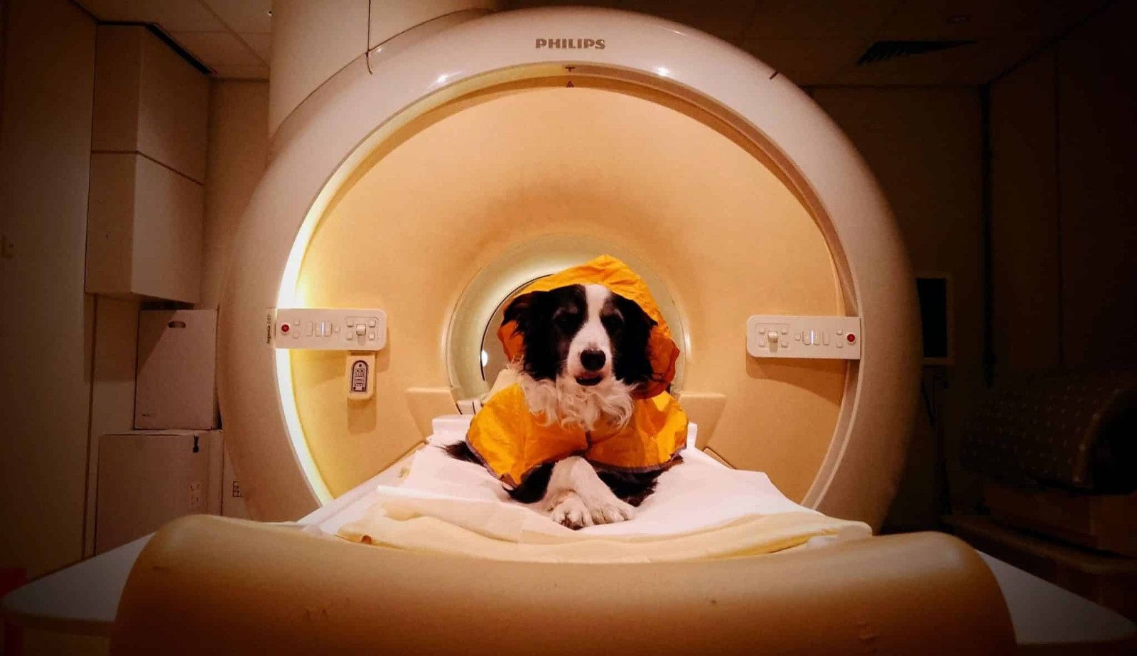
The research team designed two experiments. To avoid the influence of dogs' emotional responses to familiar people on the results, facial photos of strangers were used in the experiments. The first experiment included 8 dogs aged 18 to 53 months, including 6 Border Collies, 1 Labrador Retriever, and 1 Golden Retriever. The dogs remained in a stationary position in the scanner and could leave the device at any time. The experiment first compared happy and neutral human face images, with each photo presented for 7 seconds, followed by a 12.25-second blank screen interval, and each dog underwent 5 fMRI scans.
The results showed that when dogs viewed happy expressions, a large area of the right temporal cortex in the brain exhibited more pronounced blood oxygen level-dependent signals (BOLD signals), extending to the caudate nucleus, with a related signal cluster volume of 25,327 cubic millimeters. The researchers then further determined whether this region was sensitive only to happy expressions or responded to all emotional human faces.
The second experiment included 12 dogs, 6 of which had participated in the first phase of the experiment. The researchers used machine learning methods to analyze the activity patterns of the brain regions identified in the first experiment and had a classifier determine the type of facial expression the dogs were viewing. The results showed that this brain region could statistically distinguish happy expressions from three types of negative expressions: the average classification accuracy for happy versus sad was 69.8%, happy versus angry was 60.9%, and happy versus fearful was 63.5%. However, when comparing among the three negative expressions—sad, angry, and fearful—the classification accuracy did not exceed random levels.
To further identify differences, the researchers conducted a whole-brain analysis. The results found that activity patterns in the right anterior lateral gyrus could distinguish sad from fearful expressions; the right middle lateral gyrus and left splenial gyrus could distinguish angry from fearful expressions. For sad versus angry expressions, no statistically significant differences were found. The researchers believe that different regions in the canine brain respond differently to certain human negative emotions, with fearful expressions potentially having a stronger arousal effect or specific behavioral significance for dogs.
The study authors stated that this is the first study to provide neuroimaging evidence suggesting that canine brains can perceive certain human negative emotions. However, the study also has limitations: only 12 dogs participated in the machine learning experiment, a relatively small sample size; most of the subject dogs were Border Collies, so the results cannot be fully generalized to all dog breeds; additionally, the experiment used static photos, whereas in real life, dogs typically understand human emotions through a combination of expressions, movements, sounds, and other cues.
Disclaimer: Information republished from partner media, institutions or other websites is provided for reference and communication purposes only. It does not imply endorsement of its views or verification of its accuracy. Please contact us if any content infringes rights or requires correction.