
ORNL Neutron Imaging Technology Reveals Healed Rib Fracture in Tyrannosaurus Rex Fossil
 Scan to share
Neutron imaging technology at the U.S. Department of Energy's Oak Ridge National Laboratory (ORNL) has helped researchers from the University of Regina in Canada and other institutions observe rare healing signs inside the fossilized bones of a Tyrannosaurus rex, providing new insights into ancient life from approximately 66 million years ago.
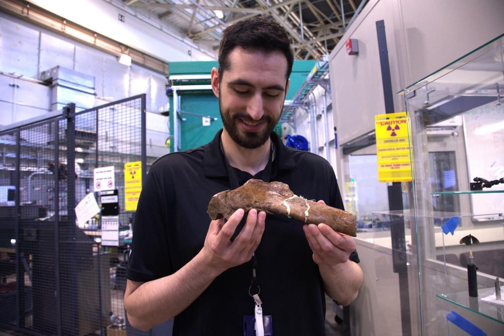
The subject of this study is a fractured rib from "Scotty," the largest Tyrannosaurus rex skeleton ever discovered. Researchers found that this rib preserved a wound-healing process rarely seen in the fossil record: after the fracture occurred, iron-rich blood entered the injured area and formed a network of blood vessels to promote healing; however, before the rib had fully healed, "Scotty" died and was preserved in a salt marsh environment, which slowed the decomposition of the remains and allowed some fragile structures to be preserved.
Mauricio Barbi, a physics professor at the University of Regina, stated that the rib of "Scotty" contains extensive mineralized vascular networks, a phenomenon not commonly observed in fossils previously. The project was co-led by Jerit Mitchell, a doctoral candidate at the University of Rochester. The research team believes that each fossil is like a tiny snapshot left from the past, and by piecing together these clues, we can better understand ancient organisms and their living environments.
The research dates back to 2020, when Mitchell first discovered vascular traces in the rib of "Scotty" using micro-CT scanning technology at the Canadian Light Source. Subsequently, the team combined synchrotron radiation imaging and microscopy techniques to study bone healing and fossilized tissue at the cellular scale. To further search for evidence of preserved soft tissue, the researchers sent samples to Oak Ridge National Laboratory for non-destructive imaging using the Multimodal Advanced Radiography Station (MARS) at the High Flux Isotope Reactor (HFIR) and the VENUS neutron imaging beamline at the Spallation Neutron Source (SNS).
Neutron imaging and X-ray imaging offer complementary advantages. X-rays are better at revealing heavier elements and dense structures, while neutrons are more sensitive to light elements such as hydrogen, enabling the detection of information that other methods may struggle to obtain. The research team used cold neutrons produced at MARS for high-resolution imaging of smaller bones, amber, and fossilized scales, and used high-energy neutrons from VENUS for three-dimensional imaging of large bone samples, including the rib of "Scotty."
ORNL instrument scientists noted that neutrons are typically used for battery or advanced materials research, but they also hold unique value in exploring ancient life. The research team plans to continue analyzing the data already obtained and expand the study to more fossil samples, comparing healing patterns across different species, while combining neutron imaging and X-ray techniques to investigate pathological features in fossils and their differences from pathologies in modern species.
Disclaimer: Information republished from partner media, institutions or other websites is provided for reference and communication purposes only. It does not imply endorsement of its views or verification of its accuracy. Please contact us if any content infringes rights or requires correction.