
Penza State University in Russia Develops Training Simulator for X-ray Image Defect Recognition
 Scan to share
Researchers at Penza State University in Russia have developed an interactive training simulator for identifying defects in X-ray images, aimed at helping medical students and residents more accurately assess abnormalities in medical images and understand the physical causes behind different image defects.
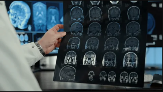
X-ray image quality directly affects the efficiency of pathological change detection and clinical diagnostic judgment. Factors such as equipment condition, exposure parameters, radiation dose, and quantum noise levels can all lead to degraded image quality. Therefore, radiologic technologists and radiologists need not only to identify lesions in images, but also to assess image quality and determine whether defects such as low contrast, noise, blur, and artifacts may affect diagnosis.
According to reports, the simulator employs a modular architecture consisting of three components: a database, an indicator calculation subsystem, and a training and testing subsystem. The database is used to store X-ray images, quality indicators, and related information; the indicator calculation subsystem can load images, calculate quantitative indicators such as effective signal-to-noise ratio, image gradient, and the difference between maximum and minimum brightness, and generate charts and reports; the training and testing subsystem provides training tasks to students through a web interface.
Leonid Krivonogov, Professor of the Department of Medical Cybernetics and Informatics at Penza State University and Doctor of Engineering Sciences, stated that all indicators in the system are based on rigorous mathematical formulas and are calculated directly from pixel data, making the results reproducible and objective.
During use, radiologists first upload images to the database, and the system automatically calculates and saves quality indicators for each image. Experts then select the highest-quality image as a reference image. The training system generates distorted images based on the reference image to simulate real defects such as low contrast, noise, blur, and artifacts. Students can view the reference and distorted images simultaneously and correlate visual features with quantitative indicators through system feedback.
The development team has completed an evaluation of 200 test cases using the simulator. Researchers selected 5 reference images of the chest, extremities, and abdomen, and generated 10 distorted images for each of the 4 defect types. The developers stated that, with precisely set indicator thresholds, the system correctly identified defect types in all test cases.
The researchers believe that the simulator can help trainees develop the visual ability to identify defects in X-ray images and further understand the causes of such defects. Students and residents can intuitively see how factors such as improper exposure parameters, patient movement, or post-processing distortions are reflected in images. The system also supports disabling hints and automatic scoring functions, allowing instructors to objectively assess student mastery and adjust teaching arrangements accordingly. At present, students at the Medical Institute of Penza State University have begun using this interactive training simulator for relevant training.
Disclaimer: Information republished from partner media, institutions or other websites is provided for reference and communication purposes only. It does not imply endorsement of its views or verification of its accuracy. Please contact us if any content infringes rights or requires correction.