
Japanese Team Achieves Femtosecond Soft X-ray Hyperspectral Microscopy, Simultaneously Acquiring Spatial and Energy Information with Single Pulse
 Scan to share
On August 21, the Japan Science and Technology Agency, RIKEN, and the University of Tokyo announced that a research team comprising Assistant Professor Yoko Takeo and Associate Professor Takashi Kimura from the Institute for Solid State Physics at the University of Tokyo, Assistant Professor Satoru Egawa from the Research Center for Advanced Science and Technology at the University of Tokyo, and Team Leader Makina Yabashi from the RIKEN SPring-8 Center, among others, successfully developed and implemented a femtosecond soft X-ray hyperspectral microscope. The findings were published on August 20 in the U.S. scientific journal *Optica*.
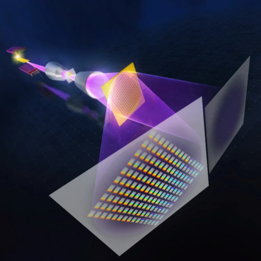
This technology utilizes a single soft X-ray pulse from an X-ray free-electron laser (XFEL) to simultaneously acquire soft X-ray spectra transmitted through different positions of a sample, enabling the synchronous acquisition of spatial and energy information in a single shot. The research team stated that this method overcomes the conventional limitation where spatial resolution and energy resolution are difficult to achieve simultaneously, providing a new observational tool for the analysis of rapidly and non-uniformly changing samples.
In terms of system design, the research team combined an aplanatic Wolter mirror microscope with a newly fabricated multi-aperture diffraction grating. The Wolter mirror magnifies soft X-ray images over a wide energy range, while the multi-aperture diffraction grating integrates 417 microscopic spectral elements, dividing the magnified two-dimensional image into multiple spatial channels and synchronously recording spectral information from each channel on a two-dimensional detector. With this configuration, the researchers were able to overlay "absorption information at different positions and different energies" onto the microscopic image, enabling analysis of spatial differences in internal composition and chemical states within the sample.
To validate the technology, the research team conducted experiments at the soft X-ray beamline BL1 of the SACLA X-ray free-electron laser facility in Harima Science Garden City, Hyogo Prefecture. The results demonstrated that the system could visualize spectral fluctuations and spatial inhomogeneities of the XFEL beam across different pulses. The researchers also used a silicon nitride thin film as a sample, capturing transmission spectral changes caused by the presence or absence of the sample near the silicon absorption edge using a single pulse with a pulse duration of less than 100 femtoseconds.
The research team believes that this achievement can serve as a foundational technology for high-speed soft X-ray spectral imaging without the need for scanning. In the future, it is expected to be applied to localized analysis of materials and devices, studies of catalytic reactions and degradation processes, chemical-state imaging of biological samples, and beam diagnostics in XFEL experiments. Subsequent research will focus on improving the bandwidth of the light source and the performance of detectors and spectral elements to further advance femtosecond-scale spatiotemporal imaging capabilities.
Disclaimer: Information republished from partner media, institutions or other websites is provided for reference and communication purposes only. It does not imply endorsement of its views or verification of its accuracy. Please contact us if any content infringes rights or requires correction.