
Russian scientists use 3D-printed tungsten grid to improve X-ray imaging quality, potentially reducing patient radiation exposure
 Scan to share
The press service of the Russian Ministry of Education and Science recently stated that a Russian research team has used 3D printing technology to manufacture a tungsten-based anti-scatter grid for X-ray machines, with partition thickness only one-third that of similar products. The researchers believe this achievement could help reduce patient radiation exposure during X-ray examinations while maintaining medical image quality.
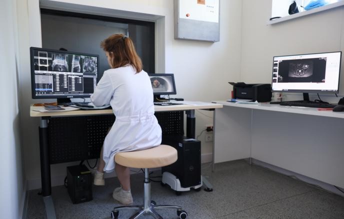
During X-ray imaging, some rays scatter after passing through human tissue, creating background noise that weakens image contrast. Clinical equipment typically suppresses such interference through anti-scatter grids. Traditional anti-scatter grids are mostly made of lead, but lead has low mechanical strength and imposes high requirements on processing techniques, often resulting in thicker structures that also absorb some of the effective rays used for imaging.
Researchers from the National University of Science and Technology MISIS (NUST MISIS), the Institute for Nuclear Research of the Russian Academy of Sciences, the Kurchatov National Research Center, and Moscow Polytechnic University proposed using selective laser melting technology with micron-scale tungsten powder to fabricate thin-walled grid structures. The high density of tungsten allows the partitions to be made thinner; at the same time, the team designed the grid channels in a converging geometry aligned along the X-ray beam direction to more effectively suppress scattered radiation while minimizing impact on the primary beam.
Stanislav Chernyshikhin, head of the Additive Manufacturing Laboratory at NUST MISIS, stated that the team has tested the grid under conditions close to actual diagnostics. They performed X-ray imaging on a model of the human pelvic region and placed test markers requiring identification in the images. The results showed that without the anti-scatter grid, the images were predominantly covered by background noise, making the marker shadows nearly indistinguishable; with the tungsten grid, the relevant markers were clearly visible.
The researchers also noted that the anti-scatter grid itself may leave shadows in X-ray images. Igor Dyachkov, an engineer at the Center for Interaction and Cooperation with Large Scientific Infrastructure at NUST MISIS, stated that the team used fast Fourier transform methods for digital image processing and successfully removed the grid shadows without losing diagnostic information. This processing method can also be applied to traditional lead-based anti-scatter filters.
The research team pointed out that in examinations of dense body regions, scattered radiation has a more pronounced effect on image quality, so finer tungsten-based anti-scatter grids may offer greater value in such clinical scenarios. The relevant research results have been published in the Bulletin of the Lebedev Physics Institute.
Disclaimer: Information republished from partner media, institutions or other websites is provided for reference and communication purposes only. It does not imply endorsement of its views or verification of its accuracy. Please contact us if any content infringes rights or requires correction.