
Japanese Research Uses Tau PET to Reveal Association with Symptom Progression in Progressive Supranuclear Palsy
 Scan to share
A research team at the National Institutes for Quantum Science and Technology (QST) in Japan recently announced findings from a study on progressive supranuclear palsy (PSP). Using their self-developed tau PET imaging technology, the researchers conducted follow-up scans on the same group of PSP patients at approximately one-year intervals, confirming for the first time in living brains that the faster tau protein accumulates in the globus pallidus region deep within the brain, the faster patient symptoms progress. The findings were published on September 5, 2026, in the international neurology journal *Movement Disorders*.
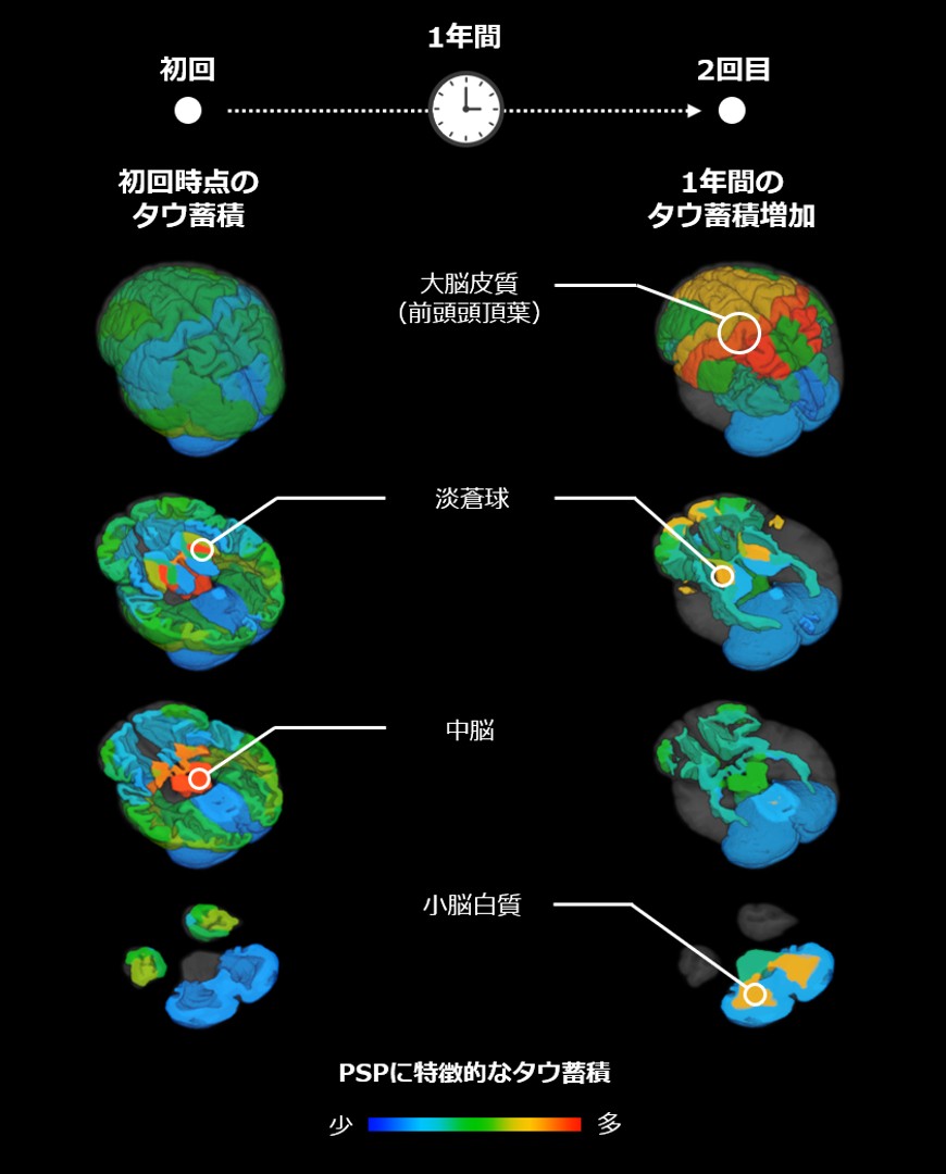
Characteristic tau protein accumulation regions in PSP
Progressive supranuclear palsy is a progressive neurodegenerative disease occurring in middle-aged and older adults, with common manifestations including increased risk of falls, difficulty with movement, eye movement disorders, postural instability, and cognitive dysfunction. There is currently no therapy that can cure or halt disease progression; therefore, establishing biomarkers that can objectively reflect disease changes and treatment efficacy has been a critical issue in drug development.
Previous autopsy studies have shown that abnormal tau protein accumulation in the brains of PSP patients is an important pathological feature, particularly concentrated in deep brain regions such as the globus pallidus and midbrain. However, autopsy samples cannot be used for long-term dynamic observation, and how tau protein accumulation changes over time and how it corresponds to symptom progression had previously remained unclear. In recent years, positron emission tomography (PET) has made it possible to observe tau protein in living brains, but existing studies have mainly shown that tau protein increases over time without sufficiently demonstrating a direct association between these changes and symptom worsening.
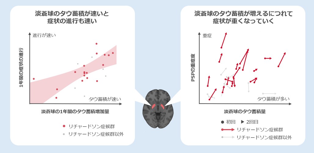
Relationship between changes in tau protein accumulation in the globus pallidus over time and symptom progression
In this study, the research team used the PET radiopharmaceutical 18F-PM-PBB3 developed by QST, namely florzolotau (18F), to perform two PET examinations on 26 PSP patients and 10 healthy controls, conducted at baseline and approximately one year later. The researchers combined 57 brain regions identified by MRI and performed multivariate analysis of PET signal changes to identify PSP-related tau protein accumulation regions and their spread over time.
The results showed that at baseline, tau protein in PSP patients was primarily accumulated in subcortical regions such as the globus pallidus and midbrain, consistent with previous reports. Follow-up imaging at one year showed further increases in tau protein accumulation in the globus pallidus, along with marked increases in tau accumulation in the frontoparietal lobes and cerebellar white matter, suggesting that during the course of PSP, tau protein may spread from deep brain regions to the cerebral cortex and cerebellar-related areas.
The research team further focused on Richardson's syndrome, the most typical and common subtype of PSP. This subtype typically progresses rapidly, and the 18 patients with this subtype included in this study showed significant increases in PSP rating scale scores within one year. The analysis found that changes in tau protein accumulation in the globus pallidus showed the strongest correlation with the degree of symptom progression; that is, the faster tau protein accumulated in this region, the more pronounced the worsening of patient symptoms. Analyses of individual patients likewise showed that globus pallidus tau protein accumulation can reflect the course of PSP from mild to severe stages.
The researchers believe that serial tau PET imaging has the potential to serve as a novel imaging biomarker for assessing PSP disease progression, and also provides a basis for tau-targeted therapeutic strategies: if future therapies can inhibit tau protein accumulation, it may be possible to slow symptom progression. Next steps still require longer-term follow-up in a larger number of patients to validate the relationship between changes in tau protein accumulation and overall symptom progression and treatment response.
The imaging assessment method established in this study may not only be used for disease monitoring and clinical trial efficacy evaluation in PSP, but may also be extended to research on other tau protein-related neurodegenerative diseases and dementias, providing new technical support for the diagnosis and therapeutic development of related diseases.
Disclaimer: Information republished from partner media, institutions or other websites is provided for reference and communication purposes only. It does not imply endorsement of its views or verification of its accuracy. Please contact us if any content infringes rights or requires correction.