
Russian research team reveals submicroscopic pore structure of chernozem using ion beam scanning electron microscopy
 Scan to share
Researchers at the Moscow Institute of Physics and Technology and their collaborators have recently used focused ion beam scanning electron microscopy (FIB-SEM) to perform detailed characterization of the microstructure of chernozem soil samples. The study shows that this technique can identify pores smaller than 1 micron, pore morphology, and specific organic components in soil, providing a new observational tool for understanding the mechanisms of chernozem fertility formation and maintenance. The research was supported by a grant from the Russian Science Foundation and published in the 17th series "Soil Science" of the Moscow University Bulletin.
Soil science research typically requires extending from the field scale to the microscopic scale. Computed tomography (CT), which can reconstruct the three-dimensional structure of soil without damaging samples, has been widely used to observe internal pore networks in soil. However, CT has resolution limitations, primarily revealing larger pore channels, while finer pores formed by complex soil processes are difficult to fully resolve.
To address this shortcoming, scientists from the Moscow Institute of Physics and Technology, Lomonosov Moscow State University, Dokuchaev Soil Science Institute, and the Schmidt Institute of Physics of the Earth of the Russian Academy of Sciences applied FIB-SEM to study chernozem samples from the Central Chernozem State Nature Reserve. This method uses a beam of ionized gallium atoms to sequentially slice soil samples, exposing internal structures, which are then imaged at high resolution by a scanning electron microscope—akin to using an "ion scalpel" combined with high-magnification microscopy to observe internal soil details.
The researchers first used a CT scanner to build a three-dimensional model of soil aggregates at a resolution of 1 micron, distinguishing pores from solid components through image segmentation algorithms. They then used FIB-SEM to cut micro-trenches in the soil, obtaining images with resolutions up to 4 nanometers. Because the sample surface contains complex structures such as clay and organic matter, the research team manually traced pore boundaries to improve recognition accuracy.
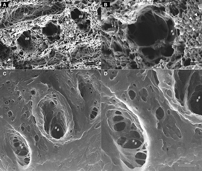
The results showed that the two techniques provide complementary information on soil structure. CT offers a larger-scale view of the overall structure, while FIB-SEM can detect features as small as 0.075 microns, or 75 nanometers, capturing both nanopores and larger pores simultaneously. The study also found that FIB-SEM can clearly distinguish organic matter within soil aggregates. In the images, this organic matter appears as uniformly gray spots ranging from 1 to 10 microns in size, whereas it is difficult to differentiate from the background in CT images.
In terms of pore morphology, soil pores in CT images are mostly circular, while FIB-SEM revealed numerous cracks as well as elongated, jagged pores within the samples. These microcracks form the microscopic pore network of chernozem, helping to explain the structural basis for its water retention and aeration properties.
The research team also noted that FIB-SEM has a narrow field of view, approximately 30×50 microns, and therefore cannot fully replace CT. Combining the two methods enables the construction of a more complete three-dimensional soil image, from large-scale structures down to nanoscale pores and organic particles, providing support for further studies of soil structure evolution, filtration properties, and aggregate formation processes. Researchers at the Moscow Institute of Physics and Technology stated that the team will continue to investigate the structural characteristics of organo-mineral compounds at the nanoscale and develop multiscale models of soil structure.
Disclaimer: Information republished from partner media, institutions or other websites is provided for reference and communication purposes only. It does not imply endorsement of its views or verification of its accuracy. Please contact us if any content infringes rights or requires correction.