
Search Results
Keyword:Tomo

Brookhaven National Laboratory Develops Integrated X-ray Imaging Tool for Nuclear Materials Research
A research team at the U.S. Department of Energy's Brookhaven National Laboratory has recently developed and built a new experimental apparatus that integrates four X-ray computed tomography techniques into a single instrument, enabling multiscale, comprehensive studies of next-generation nuclear reactor materials. The related technical achievements and capability demonstration have been published in the Journal of Synchrotron Radiation. The apparatus is located at the X-ray Powder Diffraction (XPD) beamline of the National Synchrotron Light Source II (NSLS-II) and was commissioned jointly by the U.S. Department of Energy Office of Nuclear Energy's Nuclear Science User Facilities program, Brookhaven National Laboratory's Nuclear Science and Security Department, and NSLS-II. Researchers said that next...
2026-09-12

Siemens Healthineers Completes PET Detector Manufacturing Expansion Project in Tennessee, USA
Siemens Healthineers recently completed the expansion and renovation project of its PET detector center in Rockford, Tennessee, USA, with a total investment of $43.6 million. The project aims to enhance PET detector manufacturing capacity to meet the growing demand for PET/CT scanning equipment. The project added 40,000 square feet of building area to the existing 97,000-square-foot campus and renovated 7,500 square feet of existing space. The Rockford facility is Siemens Healthineers' only manufacturing site globally for producing PET detectors used in its PET/CT systems. The expansion includes 36,000 square feet of dedicated manufacturing space and a 4,000-square-foot expansion of R&D facilities; meanwhile, the company also renovated...
2026-09-10

South Korea's DuChemBio to Build Country's First Radiopharmaceutical CDMO Plant
South Korean radiopharmaceutical therapy company DuChemBio will build the country's first contract development and manufacturing organization (CDMO) plant dedicated to radiopharmaceuticals, in response to growing demand for radiopharmaceuticals used in the diagnosis and treatment of cancer and neurological diseases. DuChemBio is a subsidiary of healthcare distributor Geo-Young Group. The company recently signed an investment agreement with North Jeolla Province and the city of Jeongeup to invest KRW 34.3 billion (approximately USD 25.5 million) in building a new plant in Jeongeup. The company stated that this is the largest investment since Geo-Young acquired DuChemBio. According to the plan, the new facility will integrate radioisotope production...
2026-09-08
Japanese Research Uses Tau PET to Reveal Association with Symptom Progression in Progressive Supranuclear Palsy
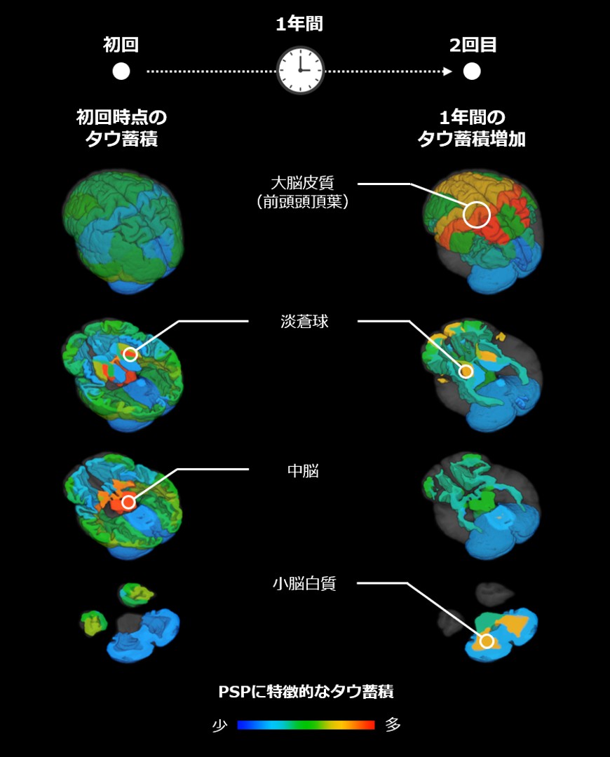
A research team at the National Institutes for Quantum Science and Technology (QST) in Japan recently announced findings from a study on progressive supranuclear palsy (PSP). Using their self-developed tau PET imaging technology, the researchers conducted follow-up scans on the same group of PSP patients at approximately one-year intervals, confirming for the first time in living brains that the faster tau protein accumulates in the globus pallidus region deep within the brain, the faster patient symptoms progress. The findings were published on September 5, 2026, in the international neurology journal *Movement Disorders*. Characteristic tau protein accumulation regions in PSP Progressive supranuclear palsy is a progressive neurodegenerative disease occurring in middle-aged and older adults, with common manifestations including increased risk of falls, difficulty with movement, eye movement disorders, postural instability, and cognitive dysfunction. There is currently no therapy that can cure or halt disease progression; therefore, establishing biomarkers that can objectively reflect disease changes and treatment efficacy has been a critical issue in drug development. Previous autopsy studies have shown that abnormal tau protein accumulation in the brains of PSP patients is an important pathological feature, particularly concentrated in deep brain regions such as the globus pallidus and midbrain. However, autopsy samples cannot be used for long-term dynamic observation, and how tau protein accumulation changes over time and how it corresponds to symptom progression had previously remained unclear. In recent years, positron emission tomography (PET) has made it possible to observe tau protein in living brains, but existing studies have mainly shown that tau protein increases over time without sufficiently demonstrating a direct association between these changes and symptom worsening. In this study, the research team used the PET radiopharmaceutical 18F-PM-PBB3 developed by QST, namely florzolotau (18F), to perform two PET examinations on 26 PSP patients and 10 healthy controls, conducted at baseline and approximately one year later. The researchers combined 57 brain regions identified by MRI and performed multivariate analysis of PET signal changes to identify PSP-related tau protein accumulation regions and their spread over time. The results showed that at baseline, tau protein in PSP patients was primarily accumulated in subcortical regions such as the globus pallidus and midbrain, consistent with previous reports. Follow-up imaging at one year showed further increases in tau protein accumulation in the globus pallidus, along with marked increases in tau accumulation in the frontoparietal lobes and cerebellar white matter, suggesting that during the course of PSP, tau protein may spread from deep brain regions to the cerebral cortex and cerebellar-related areas. The research team further focused on Richardson's syndrome, the most typical and common subtype of PSP. This subtype typically progresses rapidly, and the 18 patients with this subtype included in this study showed significant increases in PSP rating scale scores within one year. The analysis found that changes in tau protein accumulation in the globus pallidus showed the strongest correlation with the degree of symptom progression; that is, the faster tau protein accumulated in this region, the more pronounced the worsening of patient symptoms. Analyses of individual patients likewise showed that globus pallidus tau protein accumulation can reflect the course of PSP from mild to severe stages. The researchers believe that serial tau PET imaging has the potential to serve as a novel imaging biomarker for assessing PSP disease progression, and also provides a basis for tau-targeted therapeutic strategies: if future therapies can inhibit tau protein accumulation, it may be possible to slow symptom progression. Next steps still require longer-term follow-up in a larger number of patients to validate the relationship between changes in tau protein accumulation and overall symptom progression and treatment response. The imaging assessment method established in this study may not only be used for disease monitoring and clinical trial efficacy evaluation in PSP, but may also be extended to research on other tau protein-related neurodegenerative diseases and dementias, providing new technical support for the diagnosis and therapeutic development of related diseases.
2026-09-07
Domestically Developed "Palm-Sized CT" Debuts: 6 kg Micro X-ray CT Enables 3D Non-Destructive Testing
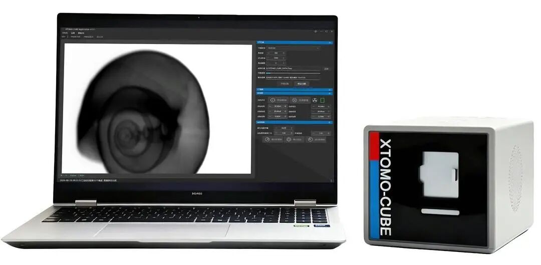
The XTOMO-CUBE series micro-CT, independently developed by Ruiying Detection Technology (Jinan) Co., Ltd., recently made its debut. With dimensions of 140×132×220 mm and a total weight of approximately 6 kg, the device can be held in one hand and is dubbed the "palm-sized CT." It can scan small-sized samples such as chicken bones, snail shells, and capsule tablets, revealing their internal three-dimensional structures. According to reports, CT stands for computed tomography technology. Compared with traditional CT equipment, the XTOMO-CUBE integrates key modules such as a miniaturized X-ray source, rotation stage, and detector into a compact body, paired with fully self-developed software capable of completing processes including projection data acquisition, CT image reconstruction, and 3D rendering. The...
2026-09-04
Cubresa Brain PET Scanner Receives FDA 510(k) Clearance, Integrates with Existing PET/MR Systems
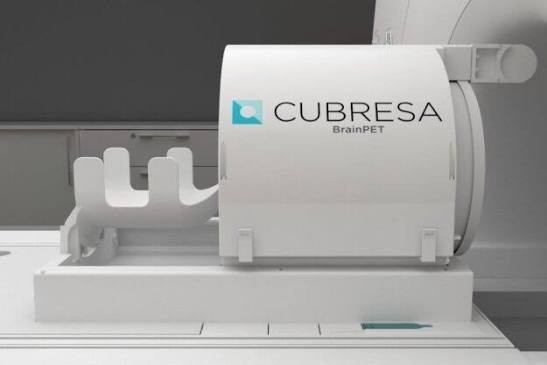
Molecular imaging technology company Cubresa Inc. announced on August 20 that its Cubresa BrainPET™ scanner has received 510(k) clearance from the U.S. Food and Drug Administration (FDA). According to the company, the Cubresa BrainPET™ is a positron emission tomography (PET) device specifically designed for brain imaging and can be integrated into existing Siemens Biograph mMR MR/PET systems. The device enables healthcare institutions to perform simultaneous PET/MR brain imaging on existing MRI infrastructure, capturing molecular and anatomical information without the need to purchase a separate whole-body hybrid imaging system. Cubresa founder and CEO James ...
2026-08-22
Global PET Market Expected to Reach $2.81 Billion by 2035, PET-CT Remains Dominant
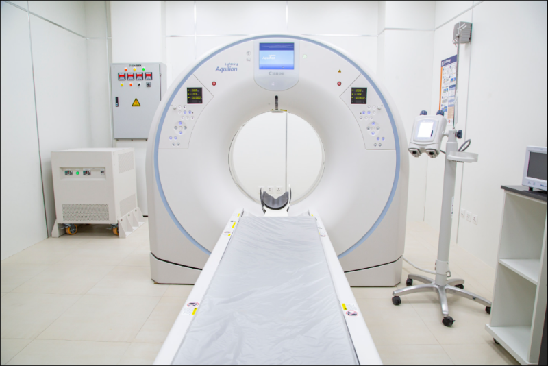
A market research report shows that the global Positron Emission Tomography (PET) market size is expected to increase from $1.86 billion in 2025 to $2.81 billion by 2035, with a compound annual growth rate (CAGR) of 4.26% from 2026 to 2035. Increased demand for cancer diagnosis, expanded application of hybrid imaging technologies such as PET-CT and PET-MRI, and continued development of molecular imaging technologies are considered major factors driving market growth. The report notes that PET scans are being increasingly used in scenarios such as cancer detection and treatment evaluation, cardiac disease assessment, and diagnosis of neurological and psychiatric disorders. Meanwhile, population aging, rising healthcare expenditures, and growing demand for early screening are also driving...
2026-08-23

Decoding the Clinical-Radiological Paradox: Brain Network Mechanisms in Spinocerebellar Ataxia Type 3 Revealed
On August 17, the team of Professor Liu Chen and Professor Wang Jian from the Department of Radiology at Southwest Hospital of Army Medical University, in collaboration with Professor Long Zhiliang's team from the Faculty of Psychology at Southwest University, systematically revealed for the first time the micro-level molecular pathological mechanisms underlying macroscopic brain network decoupling in patients with spinocerebellar ataxia type 3 (SCA3) by integrating multimodal magnetic resonance imaging technology with high-resolution micro-biological atlases. This provides a novel perspective and objective imaging targets for resolving the clinical-radiological dissociation challenge in clinical diagnosis and treatment. The findings were published in the internationally leading journal in the field of movement disorders under the title "Putaminal Structure-Function Decoupling as an Early Candidate Imaging Biomarker for Motor Severity in Spinocerebellar Ataxia Type 3."
2026-08-18

World's first laser plasma accelerator-driven high-resolution muon imaging experiment to launch in Romania
Ideon Technologies, a global subsurface detection muon tomography company, is participating in an international research consortium to conduct the world's first high-resolution muon imaging experiment driven entirely by a laser plasma accelerator. The experiment aims to use a compact accelerator to produce muon beams on demand for non-destructive high-resolution imaging, with applications targeting mineral production, critical infrastructure, cargo inspection, medical imaging, semiconductors, nuclear security, and space radiation testing. Consortium members include the University of Texas at Austin, the Extreme Light Infrastructure - Nuclear Physics (ELI-NP) facility team, Heinrich Heine University Düsseldorf, Helmholtz-Zentrum Dresden-Rossendorf, ELI-Beamlines, and Tau Systems.
2026-08-18

Neutron and X-ray tomography reveal hydrogen-rich regions in Martian meteorite "Black Beauty"
An international team of researchers from Denmark, Sweden, and Switzerland recently conducted a three-dimensional non-destructive analysis of a slice of the Martian meteorite NWA 7034 "Black Beauty" using neutron tomography and X-ray tomography, discovering macroscopic hydrogen-rich regions within it. The study suggests that these hydrogen-rich features provide evidence of early water-rock interactions on Mars. The findings have been published in Geophysical Research Letters. Black Beauty is a brecciated meteorite from Mars that was ejected into space by an impact event and eventually landed in the Sahara Desert in Morocco. Weighing approximately 320 grams, the meteorite contains material up to 4.48 billion years old, making it one of the oldest known Martian crustal fragments...
2026-08-17

Russia's MEPhI Receives Over 1 Billion Rubles in Funding for Two Domestic Scientific Instrument Projects
The Russian Ministry of Science and Higher Education will allocate 2.81 billion rubles to support the development of eight types of domestically produced scientific research equipment. According to the results of an additional competitive selection, the funds are planned to be disbursed from 2026 to 2028, with six universities and research institutions set to receive support. Among them, the National Research Nuclear University MEPhI (Moscow Engineering Physics Institute) will receive the largest share, with two projects totaling slightly over 1 billion rubles in funding. Under the arrangement, MEPhI will receive 591 million rubles for the development of a tandem quadrupole time-of-flight mass spectrometer (QTOF MS), and an additional 433 million rubles for the atom probe tomography project. Both projects have previously been approved by the Russian Academy of Sciences and included in...
2026-08-12

South Korea to Launch Real-Time Query System for CT and MRI Records in December to Reduce Duplicate Tests for Patients Transferring Hospitals
The Health Insurance Review and Assessment Service (HIRA) of South Korea announced on August 10 that it has established the "Appropriate Medical Utilization Review Committee" and, at its first meeting, included computed tomography (CT) and magnetic resonance imaging (MRI) examinations in the review scope of the real-time medical utilization management program. According to the plan, the program will be managed through the "Medical Care Benefit Details Confirmation System" following approval by the Minister of Health and Welfare. The system, built pursuant to Article 41-6 of the revised National Health Insurance Act, is expected to officially operate starting December 24. Once the system is in place, medical personnel will be able to query in real time the timing and relevant records of patients' CT and MRI examinations at other medical institutions before the patients undergo examinations, thereby reducing...
2026-08-11

Morehead State University Receives $50,000 Grant to Support Nuclear Medicine Technology Bachelor's Program
Morehead State University in the United States recently received a $50,000 grant to strengthen its Bachelor of Science in Nuclear Medicine Technology program, supporting the university's efforts to train medical imaging professionals for Kentucky's healthcare system. The funding was provided by the Kentucky Board of Medical Imaging and Radiation Therapy and administered by the Kentucky Council on Postsecondary Education. The project funds were awarded through a competitive bidding process, with a focus on supporting nuclear medicine technology education programs in Kentucky, aiming to increase the number of qualified professionals entering the state's healthcare industry. The newly established nuclear medicine technology undergraduate program under Morehead State University's Department of Imaging Sciences will prepare students in the field of medical imaging...
2026-08-11
Russian research team reveals submicroscopic pore structure of chernozem using ion beam scanning electron microscopy
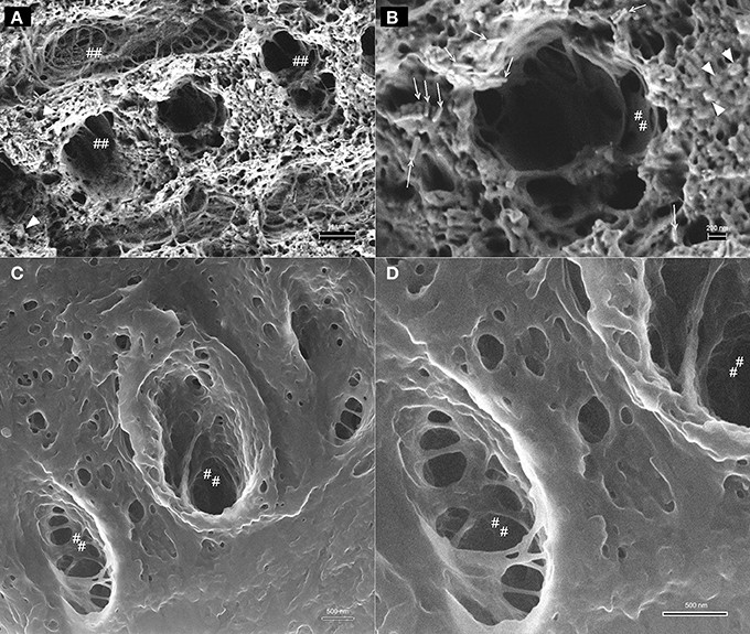
Researchers at the Moscow Institute of Physics and Technology and their collaborators have recently used focused ion beam scanning electron microscopy (FIB-SEM) to perform detailed characterization of the microstructure of chernozem soil samples. The study shows that this technique can identify pores smaller than 1 micron, pore morphology, and specific organic components in soil, providing a new observational tool for understanding the mechanisms of chernozem fertility formation and maintenance. The research was supported by a grant from the Russian Science Foundation and published in the 17th series "Soil Science" of the Moscow University Bulletin. Soil science research typically requires extending from the field scale to the microscopic scale. Computed tomography (CT) has been widely used to...
2026-08-03
U.S. Explores New Methods for Lunar Ice Detection Using Synchrotron X-ray Microtomography and Seismic Models
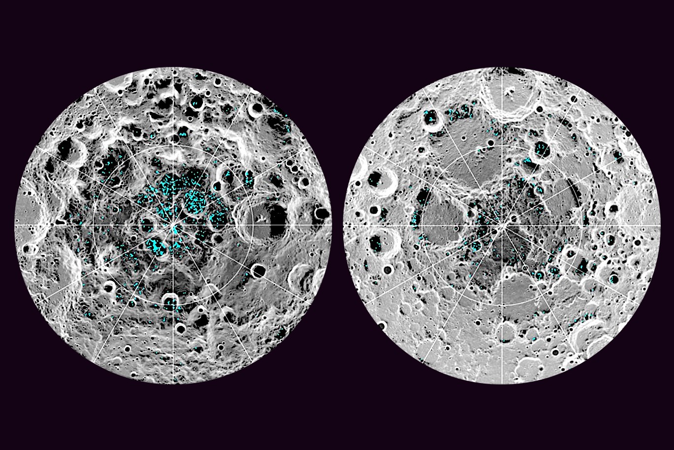
On July 31, researchers at Lawrence Berkeley National Laboratory, in collaboration with scientists from the University of Maryland and the University of Hawaii, proposed a new method for locating subsurface ice deposits on the Moon using seismic waves. The findings, published in the journal Science Advances, are expected to provide a reference for interpreting detection data and locating water resources in future lunar landing missions. The research focuses on how subsurface lunar materials respond to seismic waves. The team developed computational models to predict the behavior of lunar subsurface media of varying compositions under seismic wave excitation. Experimental results showed differences in seismic response between ice-bearing and dry rocks, indicating that seismological methods have the potential to identify ice layers buried beneath lunar regolith and rock.
2026-08-01